What Regeneration Means
Regeneration is the rebuilding of missing or damaged tissue after an injury. The result may be a complete body region, a single organ, or a replacement that works but does not copy the original perfectly. A planarian flatworm can restore a head from a small body fragment, while an axolotl can regrow a limb with bone, muscle, nerves, blood vessels, and skin. Those examples sit at opposite ends of a broad biological spectrum.
Speed and scale differ by species. A zebrafish caudal fin can finish a full regeneration cycle in about two weeks under laboratory conditions, with wound coverage beginning within hours. A recent planarian study reports new heads forming within 20 days in its experimental animals. These figures are guides, not promises: temperature, age, nutrition, wound size, genetics, and stress all influence the schedule.
Regrowth does not mean an animal is indestructible. Regeneration replaces selected structures after a controlled injury; it does not reverse every disease or repair unlimited damage. Some animals also shed and regrow parts as a normal life cycle, such as arthropods replacing a lost limb during later molts. The useful question is not simply “Can it grow back?” but which tissues return, how faithfully they return, and what signals organize the process.
Why Regrowth Fails
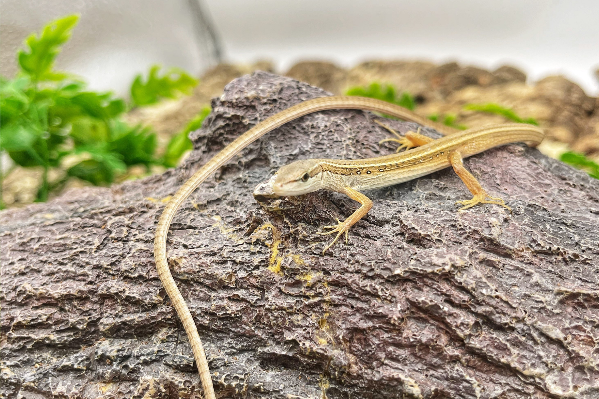
A common error is treating every replacement as a perfect duplicate. A lizard tail usually develops a cartilage tube instead of the original chain of vertebrae, so movement and strength may differ. A regenerated limb can also have altered proportions or pigmentation. The visible outline is only one measure; nerves, joints, muscles, blood supply, and behavior reveal how complete the repair is.
Another mistake is confusing healing with regeneration. Mammalian skin can close a cut, and bone can remodel after a fracture, yet a lost adult arm does not reappear. Scar formation seals a wound quickly but may limit the cell movement and signaling needed for a new structure. In species with strong regenerative capacity, wound closure is still necessary, but it is followed by a coordinated growth phase.
Laboratory conditions can create misleading expectations. A zebrafish fin is a small, accessible model with a well-studied wound surface; it is not a miniature human limb. A planarian’s stem-cell pool and body plan are unlike those of a mammal. A result from an amputated fin, tail, or worm fragment cannot be transferred directly to human treatment.
How Animals Rebuild
Start With A Clean Wound
Regrowth begins with rapid wound coverage and a local change in chemical signals. In zebrafish fin experiments, epithelial cells migrate across the cut surface within roughly 1–3 hours, and an apical epidermal cap forms by about 18–24 hours. This temporary covering is not passive wrapping: it exchanges signals with cells underneath and helps establish the regenerative environment.
For a reader examining a study, the timing of these early events is useful. Researchers often mark wound closure, cell division, and the appearance of a blastema separately. If a paper measures only a smaller wound, the result may describe healing rather than full appendage replacement.
Build A Blastema
A blastema is a temporary group of proliferating or progenitor-like cells beneath the wound surface. Axolotl limb research shows nearby connective-tissue cells can shift into a more uniform progenitor state, help reorganize the extracellular matrix, and later redifferentiate into appropriate tissues. The cells do not become an unlimited blank slate in every species; lineage restrictions and the position of the original tissue still matter.
Zebrafish fins use a related but distinct arrangement. Injury-activated fibroblasts enter the blastema, and osteoblasts can dedifferentiate before returning to bone-forming roles. The new fin is shaped by positional information, local growth signals, nerves, and the wound epidermis. This is closer to a managed construction site than a simple swelling of generic stem cells.
Use Stem Cells Wisely
Planarians rely heavily on adult stem cells called neoblasts. These cells are distributed through the body and can generate many mature cell types, including neurons, muscle, gut, and epidermis. Research has identified subgroups with different developmental tendencies, while transplantation experiments show that a single suitably potent neoblast can rescue a stem-cell-depleted animal.
That capacity explains why a fragment can reconstruct a whole animal, but it does not make the process random. Positional signals tell new cells where a head, tail, eye, or digestive region belongs. A wound must coordinate cell division with body-axis patterning, energy use, and tissue differentiation. Disrupting one signal can produce missing, misplaced, or duplicated structures.
Stop At The Right Size
Growth must end when the replacement reaches the right dimensions. Cells compare local position with the surrounding tissues through networks of molecular signals; they also respond to mechanical pressure, nutrient status, and maturation cues. Zebrafish fins usually restore their original proportions after repeated amputations, yet the experiment still depends on a healthy animal and a defined wound.
Scientists test the stopping problem by tracking cell lineages, changing gene activity, measuring cell division, and comparing regenerated anatomy with the original. A strong study reports controls, wound location, animal age, sample size, and the time point used for measurement. Those details reveal how much of the conclusion applies beyond the model organism.
Cases From Research
Imagine an educational laboratory group cutting a small section from a zebrafish caudal fin and photographing the same fish at fixed intervals. The wound closes first, a blastema forms beneath the new epidermal cover, and bony rays extend until the fin returns near its former outline. The group should record water temperature and fin position because both affect comparisons. Repeated-amputation studies show that healthy zebrafish can retain regenerative capacity across several cycles, but that finding does not mean every injury is harmless.
Now consider a second anonymized classroom scenario involving planarians. Students divide worms under approved laboratory supervision and watch eye spots, body shape, and movement emerge over days. A head-facing fragment and a tail-facing fragment develop different structures because positional information remains in the tissue. The students should not call this “instant cloning”: cell proliferation, differentiation, and pattern correction take time, and survival depends on the fragment and conditions.
Axolotl work supplies another useful comparison. After limb amputation, connective-tissue lineages contribute much of the blastema, while muscle, nerve, skin, and skeletal components follow their own lineage histories. The new limb can be highly complete, yet the result comes from a vertebrate with a specialized regenerative program. It is evidence about cellular coordination, not a ready-made recipe for human limb replacement.
Compare Regenerative Abilities
The table separates the structure that returns from the mechanism and the main limit. “High” refers to the range of tissues commonly studied in that animal, not to an unlimited capacity.
| Animal | What Regrows | Main Cell Strategy | Key Limit |
|---|---|---|---|
| Planarian | Whole body regions and organs | Pluripotent neoblasts plus body-axis signals | Fragment size and pattern errors affect survival |
| Axolotl | Limbs, tail, and several tissues | Lineage-linked cells form a blastema | Capacity varies with tissue, age, and injury |
| Zebrafish | Fins, heart, retina, and other tissues | Blastema with mostly lineage-restricted cells | A model fin is simpler than a human limb |
| Lizard | Part of the tail | Wound response and cartilage-forming growth | New tail often lacks original vertebrae |
Common Mistakes
Do not describe all regenerative animals as using the same stem cells. Planarian neoblasts, axolotl connective-tissue cells, and zebrafish fin progenitors follow different rules. A source that uses “stem cell” as a broad label may hide lineage limits, so read the methods and cell-tracing evidence.
Do not judge success from a photograph alone. Compare length, symmetry, tissue types, sensation, movement, and repeated function when the study measures them. A lizard tail that looks complete may lack the original vertebral column. A fish fin that reaches its old length may still have a temporary difference in pigmentation during repair.
Do not turn animal research into a medical promise. Regeneration studies can clarify wound signals and cell behavior, but human healing has different immune, scar, vascular, and anatomical constraints. Avoid products or procedures that claim a worm, fish, or salamander result proves a treatment works in people.
Finally, do not ignore welfare and conservation. Amputation experiments need ethical oversight, correct anesthesia where required, and suitable housing. Wild animals should never be cut or collected to recreate a classroom demonstration. Published protocols and supervised observation are safer routes to learning.
FAQ
Which animal regrows the most?
Planarians rank among the strongest examples because small fragments can rebuild missing body regions and organs. Their ability comes from abundant neoblasts and positional signals, not from a universal rule shared by all animals.
Can an axolotl regrow a full limb?
Yes. Axolotls can regenerate an adult limb with several tissue types, although the outcome depends on injury site, age, health, and experimental conditions.
Do lizards regrow their original tails?
Usually not exactly. A replacement tail commonly contains a cartilage tube rather than the original segmented vertebrae, so its flexibility and structure can differ.
How fast does a zebrafish fin grow back?
A healthy adult zebrafish can complete much of caudal-fin regeneration in about two weeks under laboratory conditions. Temperature and wound size change the schedule.
Can humans regrow lost limbs?
Humans can repair some tissues and remodel bone, but they do not naturally regenerate a lost adult limb. Animal studies explain mechanisms without proving that human limb regrowth is available.
Author's Insight
The clearest lesson is that regeneration is an organized conversation between cells, wound surfaces, body position, and time. Whole-body regeneration in a planarian is not simply a stronger version of human healing; it rests on a different body plan and stem-cell arrangement. Axolotl and zebrafish studies show that a vertebrate can reuse local cells while retaining lineage boundaries. Comparing structure, function, and mechanism together gives a more accurate picture than spectacular photographs alone.
Key Takeaways
Animals that regrow body parts use several strategies. Planarians draw on neoblasts to rebuild broad regions; axolotls form a blastema that restores a complex limb; zebrafish rebuild fins through coordinated local progenitors; and lizards replace part of a tail with a different internal skeleton. A regenerated part may look familiar without being anatomically identical.
For reliable reading, check the animal, tissue, wound size, observation period, and measurements. The strongest evidence tracks cells and tests function, while stating the limits of the model. These animals teach biology by showing how repair can be organized, but they do not erase the major differences between species.