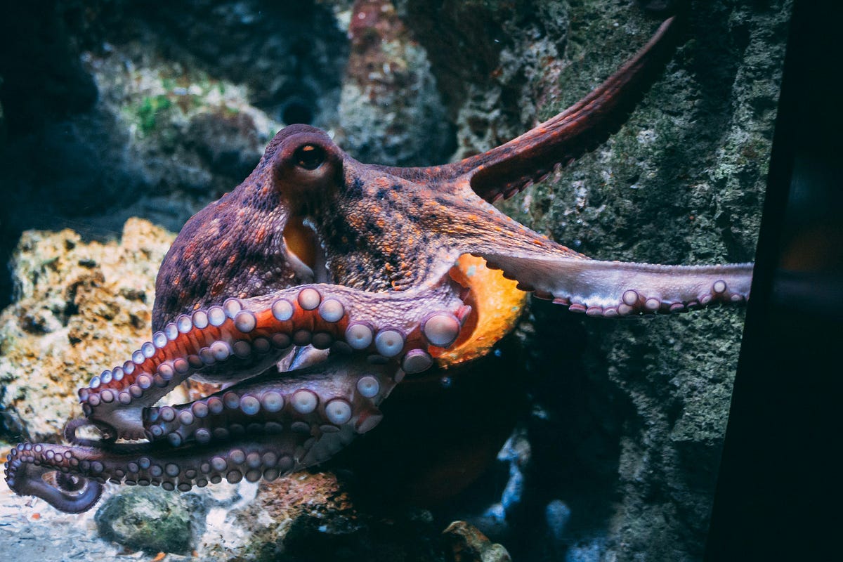
Octopus Circulation Basics
Octopuses have three hearts because their blood-oxygen path is split between the body and the gills. Two hearts pump blood to the gills, and one heart pumps oxygenated blood to the rest of the body. This design matches the animal’s need to exchange gases across gill tissue while still supporting rapid movement and hunting.
In most octopuses, blood uses hemocyanin, a copper-containing oxygen carrier. Hemocyanin gives the blood a bluish color and binds oxygen in a reversible way. One measurable detail: octopus blood can carry oxygen at relatively low partial pressures compared with many vertebrates, which helps in aquatic environments where oxygen levels vary.
Three hearts, one job.
Researchers also describe the octopus circulatory system as “closed” in the sense that blood travels through vessels rather than only through open body spaces. Even so, the system still relies on gill perfusion to load oxygen. When an octopus reduces gill flow, oxygen delivery to tissues can drop, which is one reason activity patterns matter.
Main Problems People Miss
A common misunderstanding is treating the three hearts as three independent “main pumps.” In practice, the two gill hearts and the systemic heart work in series: blood must be oxygenated in the gills before it can be distributed to organs. If you watch only one part of the cycle, the system can look confusing.
Another error is assuming the three hearts exist to “increase speed” in a simple way. The real driver is oxygen routing. The gills are the site of gas exchange, so the circulatory system must deliver blood there efficiently and then return it to the body.
People also overgeneralize from one species to all cephalopods. Some cephalopods differ in gill structure, blood chemistry, and how strongly they regulate flow. Even within octopuses, measurements can vary by temperature, size, and activity level.
Solutions And Tips
Map The Pumping Sequence
To understand the three hearts, track the sequence: two hearts send blood to the gills, then the systemic heart sends oxygenated blood to the body. This mental model prevents the “three main pumps” mistake. In practice, you can connect it to what you see: gill water flow and mantle movement often change before you notice obvious changes in behavior.
Look for gill ventilation changes.
If you keep an octopus in captivity, monitor water flow and oxygenation rather than only watching the animal. A typical aquarium oxygen test kit or dissolved oxygen probe can help you interpret behavior changes. I once saw a keeper blame “stress” for reduced activity, but a dissolved oxygen reading showed a drop after a filter adjustment; the animal’s ventilation increased afterward.
Use Oxygen Measurements, Not Guesswork
When you evaluate health or stress, measure dissolved oxygen and temperature. Oxygen binding by hemocyanin depends on conditions, and temperature changes metabolic demand. A practical target is to keep dissolved oxygen in a range appropriate for marine cephalopods; exact numbers vary by species and system, so use species-specific husbandry guidance when available.
Measure dissolved oxygen and temperature.
In research contexts, scientists often report oxygen partial pressure or dissolved oxygen alongside heart and ventilation measurements. That pairing matters because heart rate alone cannot tell you whether oxygen loading is adequate.
Interpret Behavior With Ventilation
Use mantle and jetting behavior as indirect indicators of ventilation and oxygen demand. Octopuses can jet water for locomotion, and that movement can change water flow through the mantle cavity. In practice, if an animal increases jetting and shows sustained high activity, oxygen demand rises and the gill hearts and systemic heart must keep up.
Watch mantle motion patterns.
When activity drops, gill ventilation often slows too. That does not automatically mean illness; it can reflect normal resting metabolism. Still, if the animal shows prolonged inactivity plus abnormal posture, reduced responsiveness, or persistent gill movement changes, consult a qualified aquatic veterinarian.
Account For Species And Size
Do not treat “octopus” as one physiology. Species differ in gill surface area, blood properties, and how strongly they regulate circulation. Body size also changes metabolic rate per unit mass, which affects how much oxygen delivery the hearts must support.
Compare like with like.
If you read a study, check the species name, temperature, and whether the animal was at rest or stimulated. A measurement from a small animal at one temperature may not match a larger animal in a different setting.
Recognize Limits During Low Oxygen
Three hearts cannot compensate for extremely low oxygen in the water. If dissolved oxygen drops, oxygen loading in the gills slows, and the systemic heart then distributes less oxygenated blood. In practice, you may see increased ventilation, reduced activity, or changes in feeding behavior.
Low oxygen reduces oxygen loading.
For decision support, treat oxygen problems as a system issue: check aeration, circulation, stocking density, and any recent changes to filtration or lighting schedules. If you use chemical oxygen demand removers or carbon dosing, verify that they do not create oxygen dips.
Realistic Use Case Examples
Scenario: Oxygen Drop After Filter Change
An octopus in a home aquarium becomes less active after a filter adjustment. The keeper reads “three hearts” and assumes the animal will compensate automatically. The keeper then measures dissolved oxygen and finds a temporary decline after the change, while temperature remains stable. After restoring aeration and improving water circulation, the octopus resumes normal ventilation and feeding behavior.
This scenario illustrates that heart anatomy does not override environmental oxygen limits.
Scenario: Confusing Resting vs Stress
A student records videos of an octopus that appears to “breathe” less during daytime. They interpret reduced mantle movement as illness because they focus on the idea of constant pumping. A second observation session at night shows increased ventilation during exploratory behavior, and the animal’s posture remains normal. The student concludes that ventilation varies with activity and that single-time observations can mislead.
Ventilation and behavior together give a better interpretation.
Comparison Table
| What You Notice | Most Likely Mechanism | What To Check Next | What Not To Conclude |
|---|---|---|---|
| Reduced mantle motion | Lower activity and lower ventilation demand | Compare with feeding time and observe for hours | That the three hearts are failing |
| More jetting | Higher oxygen demand and altered water flow | Check dissolved oxygen and temperature | That jetting alone diagnoses disease |
| Persistent lethargy | Possible stressor or chronic oxygen mismatch | Review water chemistry, oxygen, and recent changes | That anatomy guarantees recovery |
Checklist for interpreting “three hearts” claims:
- Identify whether the source explains gill routing, not just “three hearts.”
- Check whether it mentions hemocyanin or oxygen loading at gills.
- Look for conditions: temperature, activity state, and oxygen availability.
- Use observations that connect behavior to ventilation, not only heart count.
- If health concerns persist, seek aquatic veterinary input rather than relying on anatomy alone.
Mistakes To Avoid
One mistake is repeating the “three hearts” fact without describing the division of labor. Without the gill-heart versus systemic-heart distinction, readers cannot connect anatomy to oxygen delivery.
Another mistake is treating aquarium videos as direct evidence of heart function. Heart beats in cephalopods can be difficult to see, and water movement can mask visual cues. A claim that “the hearts are failing” based on appearance alone usually lacks support.
People also ignore environmental variables. Temperature shifts metabolic demand, and dissolved oxygen changes oxygen loading. If you do not measure those variables, you cannot interpret changes in behavior reliably.
FAQ
Do All Octopuses Have Three Hearts
Most described octopus species show a three-heart arrangement with two gill hearts and one systemic heart, but exact anatomy can vary across species and studies. If you need certainty for a specific species, use species-level references rather than general summaries.
What Do The Two Gill Hearts Do
The two gill hearts pump blood to the gills so oxygen can bind to hemocyanin in the blood. After oxygenation, blood returns to the systemic heart for distribution to body tissues.
What Does The Systemic Heart Pump
The systemic heart pumps oxygenated blood from the gills to the rest of the body through vessels. This step supports organs and muscles once oxygen loading has occurred.
Why Does Ventilation Matter For Oxygen
Ventilation moves water across gill tissue, which controls how quickly oxygen enters the blood. If water oxygen drops or ventilation slows, oxygen delivery to tissues can fall even with three hearts.
Can Heart Count Predict Endurance
Heart count alone does not predict endurance because oxygen delivery depends on dissolved oxygen, temperature, and metabolic demand. During low-oxygen conditions, performance can still drop despite the three-heart design.
Author's Insight
The three-heart design makes sense when you treat oxygen delivery as a routed process: gills load oxygen, then the systemic pump distributes it. That routing explains why gill ventilation and water oxygen concentration matter as much as anatomy. I also find that many popular explanations stop at “three hearts” and skip the oxygen pathway, which leaves readers unable to interpret behavior changes. A careful approach links anatomy, hemocyanin oxygen binding, and measurable environmental conditions like dissolved oxygen and temperature.
Key Takeaways
- Two hearts pump blood to the gills; one heart pumps oxygenated blood to the body.
- Oxygen delivery depends on gill ventilation, dissolved oxygen, and metabolic demand, not heart count alone.
- Behavior observations become more reliable when paired with measurements like temperature and dissolved oxygen.
- Species differences and study conditions can change how circulation behaves, so general claims need context.
- For health concerns in captive animals, use evidence-based husbandry checks and seek aquatic veterinary guidance when symptoms persist.